
English
Have you ever wondered how our bodies can replicate and transmit genetic information from one generation to the next? It's a fascinating process, and today we are delving into the world of DNA replication modeling. Whether you're a biology enthusiast, a genetics student, or simply curious about the intricacies of life itself, this article will take you on an enjoyable journey through the inner workings of our cells. Get ready to uncover the hidden secrets behind DNA replication as we explore them together!
1. What is DNA, the Genetic Material?
2. Discovering DNA
3. DNA Structure
4. DNA Replication Model
5. Transcribing DNA
6. Genes
7. Protein Synthesis
8. Ribonucleic Acid (RNA)
9. Regulatory Genes (Controllers)
10. Conclusion
Why were alphabet letters one of the most important things you had to learn when you entered school? Because they are the code that deciphers the secrets of the Arabic language. Similarly, cells use codes stored in their genetic material, which is made up of a chemical compound called deoxyribonucleic acid (DNA). DNA contains specific information about the growth and activity of living organisms.
Look at Figure 1, which illustrates how DNA is stored in cells that have a nucleus. When cells divide, DNA is replicated and transferred to the new cells. In this way, each new cell obtains the same information as the original cell. Therefore, every cell in your body or any other living organism must contain DNA.
This can be a good starting point for your article, and other parts can be translated in a similar manner. If you have any additional questions or need help with translating a specific part of the article, please feel free to ask.
Discovery of DNA:
Scientists discovered in the mid-1800s that the cell nucleus contains large molecules they called nucleic acids. In 1950, chemists identified the components of the nucleic acid DNA, but at that time, they couldn't create a model to describe how these components were arranged to form a DNA molecule.
In 1952, scientists Rosalind Franklin and Maurice Wilkins found that DNA is composed of two strands that have a spiral or helical shape, and using X-ray diffraction, Dr. Franklin determined that the DNA molecule has a double helix structure. In 1953, based on the work of Franklin and other scientists, James Watson and Francis Crick proposed a model for the structure of DNA.
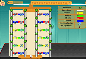
What does DNA look like? Based on the model proposed by Watson and Crick, each strand of the double helix consists of a sequence of sugar (a five-carbon sugar lacking oxygen) and phosphate groups. Meanwhile, the rungs of the ladder are made up of nitrogenous base molecules. DNA contains four types of nitrogenous bases: adenine (A), guanine (G), cytosine (C), and thymine (T). Scientists observed that the quantity of cytosine in a cell always equals the quantity of guanine, and the quantity of adenine is equal to the quantity of thymine, leading them to hypothesize that the nitrogenous bases are paired up in the DNA molecule (each base pairs with another), as depicted in the diagram, with adenine in one strand pairing with thymine in the opposite strand, and guanine pairing with cytosine. These nitrogenous base pairs are complementary, much like puzzle pieces in the structure.
When chromosomes duplicate before mitosis or meiosis, the amount of DNA inside the nucleus doubles. Watson and Crick's model illustrated how this occurs, where the DNA strands separate from each other, and new nitrogen bases pair to form a new DNA strand. The sequence of nitrogen bases in the new DNA remains the same as in the original DNA.
Most human traits, such as hair color, height, and others, rely on proteins produced by the body's cells. Proteins play a vital role in cell and tissue construction and can act as enzymes. The information used by cells to manufacture these proteins is carried by DNA. The part of DNA carried on a chromosome responsible for protein production is called a gene. Each chromosome contains hundreds of genes, as shown in Figure 3. Proteins consist of a chain of hundreds or thousands of amino acids, and the gene determines the sequence of amino acids that make up the protein. Any change in this sequence results in a different protein. But what happens to the body's cells when a protein isn't produced or a malfunction occurs in its production for some reason?
Genes are found in the nucleus. However, the process of protein synthesis takes place in ribosomes located in the cytoplasm. Therefore, the transfer of protein synthesis code from the nucleus to the ribosomes occurs through another type of nucleic acid, known as ribonucleic acid or RNA.
RNA is produced in the nucleus, and it's a copy of DNA but with some differences. Comparing the structure of DNA in Figure 4 to the structure of RNA in Figure 4, several differences are apparent, including the fact that RNA consists of a single strand while DNA has two strands. Also, RNA contains the same nitrogen bases as DNA except for thymine (T), which is replaced by uracil (U) in RNA. RNA also has a five-carbon sugar, while DNA has a five-carbon ribose sugar lacking an oxygen atom. This is why it's called ribonucleic acid. There are three types of RNA in the cell: messenger RNA (mRNA), transfer RNA (tRNA), and ribosomal RNA (rRNA). mRNA plays a crucial role in building proteins, and the process begins when RNA travels from the nucleus to the cytoplasm, where it binds with ribosomes containing rRNA scattered throughout the cell's cytoplasm.
After binding with ribosomes, the process of amino acids bonding to each other within the ribosome begins. Each nitrogen base in mRNA pairs with its counterpart in tRNA. This process continues, as shown in Figure 4. Amino acids on tRNA then link together to form a long, interconnected chain, marking the beginning of the protein chain. The code carried by mRNA determines the sequence of amino acid linkage, and once tRNA loses its amino acid, it becomes free in the cytoplasm to carry amino acids again, just as it did the first time.
You might think that all cells in a living organism manufacture the same proteins because they contain the same chromosomes and genes, but this is not the case. Each cell uses a subset of its many genes to produce the necessary proteins, and each cell uses only the genes that produce the proteins required for its specific functions. For example, muscle proteins are produced in muscle cells, not in nerve cells.
1. DNA, the genetic material, carries biological information about the growth and activities of living organisms.
2. DNA was discovered in the mid-19th century, and in subsequent decades, its structure, storage, and replication processes were understood.
3. Genes, which carry genetic information, play a fundamental role in determining the traits of living organisms and guiding protein production. This explains how proteins, a crucial part of the structure and function of living organisms, are synthesized.

© All Rights Reserved @ 2026 For VLaby - Powered By VLaby.
 English
  Arabic
  French
  German
  Indonesian
0 Comments
{{ comment.user.name }}
{{ comment.created_at }}
{{ comment.comment }}
{{ reply.user.name }}
{{ reply.created_at }}
{{ reply.comment }}
Add a Comment